Key Points
- The HotSHOT and Dipstick DNA extraction methods are quick extractions for PCR/LAMP/RPA assays, BUT they won’t produce high molecular weight DNA (e.g. for genome sequencing), or concentrate low-abundance DNA (e.g. for eDNA detection).
- The HotSHOT extraction method is extremely cheap and scalable for large numbers of samples, BUT it produces crude and dirty extracts of fragmented DNA, it may contain a lot of PCR inhibitors (substances that inhibit PCR) and its heating step requires a thermocycler.
- The Dipstick extraction method is great for samples with high concentrations of PCR inhibitors because it contains a wash step, and can be done in the field without equipment, BUT it produces only a tiny amount of DNA that goes straight to PCR, or LAMP or RPA reactions.
If you’re looking for a simple, fast DNA extraction method for PCR, you might like to consider the HotSHOT or dipstick DNA extraction methods.
- Both methods are rapid, affordable, and require no special storage or safety precautions.
- They can work with Bento Lab or any other PCR equipment.
- And we have ready-to-use kits (the HotSHOT DNA Extraction Kit, and the Dipstick DNA Extraction Kit) available in our online store.
But what applications are they suitable for? How do they differ? And which one should you choose for your application?
In this post, we discuss these two methods and their advantages and disadvantages, to help you decide if they’re appropriate methods and kits for you.
If you need purified, high-quality, high-concentration DNA, other methods may suit you better!
Before we describe and compare the HotSHOT and Dipstick DNA extraction methods, please note that they’re optimised for PCR applications (which require only a tiny fragment of target DNA for amplification) rather than applications that need total DNA extraction with minimal DNA fragmentation (such as whole genome sequencing).
In other words, they won’t produce the same quantity, quality, and purity of DNA as more expensive, time-consuming, and potentially hazardous methods. This is because of the tradeoffs that make them affordable, rapid, and safe.
If you need purified DNA extracts of high molecular weight DNA for DNA, such as when doing genome sequencing, then you will need to use other methods, for example this protocol by Jones et al. (2021).
Similarly, you’d need a different method if you need to concentrate extremely low-abundance DNA, such as when doing eDNA studies from water. For a great review of alternative options, see Rieder et al. (2024).
But if all you need is enough DNA to amplify well in a PCR, LAMP, or RPA reaction, then the HotSHOT and dipstick extraction methods may be worth considering — read on for more information!
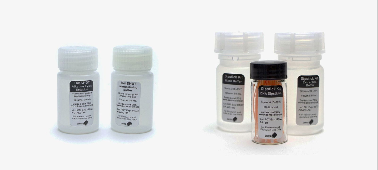
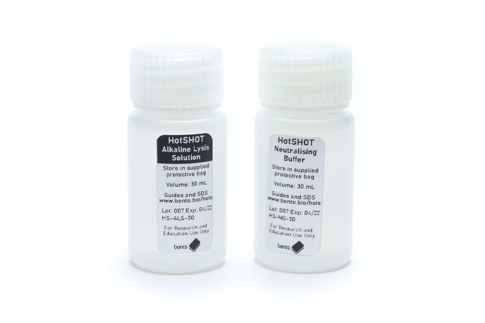
The HotSHOT DNA Extraction Method
When to consider the HotSHOT method
You should consider HotSHOT if you need a very inexpensive (<$0.10/sample), 20–30 min, scalable, and safe approach for small to large numbers of samples, that has minimal sample handling.
BUT make sure that it works for your sample types first! HotSHOT produces crude extracts that can contain PCR inhibitors (substances that inhibit PCR), and extracts may need dilution prior to PCR. The DNA will be highly fragmented, single-stranded, and less stable than other extracts, but it’s usually good enough for PCR.
HotSHOT can work great for applications like routine genotyping or high-throughput DNA barcoding — it’s very cheap and easy to scale up from a few samples to very large sample numbers. It’s also simple enough to be done by a lab robot. However, a grinding or bead-beating step may be needed for some tough sample types (e.g. seeds)
HotSHOT been used for a wide range of different sample types, including bacteria, fungi (fungal parasites in plants and diatoms), plants (leaves, seeds [with homogenisation], and pollen), invertebrates (terrestrial insects and fishbourne parasites), mammal tissues, bird feathers and bird blood, fish tissues (scales, mucus, larvae, and eggs), and even to extract viruses from human skin biopisies!
What does the HotSHOT method involve?
The HotSHOT method was first developed by Truett et al. (2000) for routine mouse tissue genotyping for medical research. The extraction method is a simple four-step process:
- Add 75 µL of Alkaline Lysis Solution to 1-2 mm3 of a sample in a 0.2 mL PCR tube.
- Incubate at 95 ºC for 20-30 minutes (depending on tissue).
- Allow to cool (ideally at 4 ºC for 5 minutes)
- Add 75 µL of Neutralising Buffer.
Between 0.5–5 µL of DNA extract can be used as DNA template per 20 µL PCR reaction.
Read published studies using our HotSHOT DNA Extraction Kit
You can read about some uses of our kit here:
- Sharrow et al. (2024). New State Record of Phytomyza ditmani Kulp (Diptera: Agromyzidae) in Arkansas. Southeastern Naturalist, 23(3), N50-N53. https://doi.org/10.1656/058.023.0315
- Ye et al. (2024). A novel in vivo genome editing doubled haploid system for Zea mays L. Nature Plants, 10(10), 1493-1501. https://doi.org/10.1038/s41477-024-01795-9
And watch our video here:

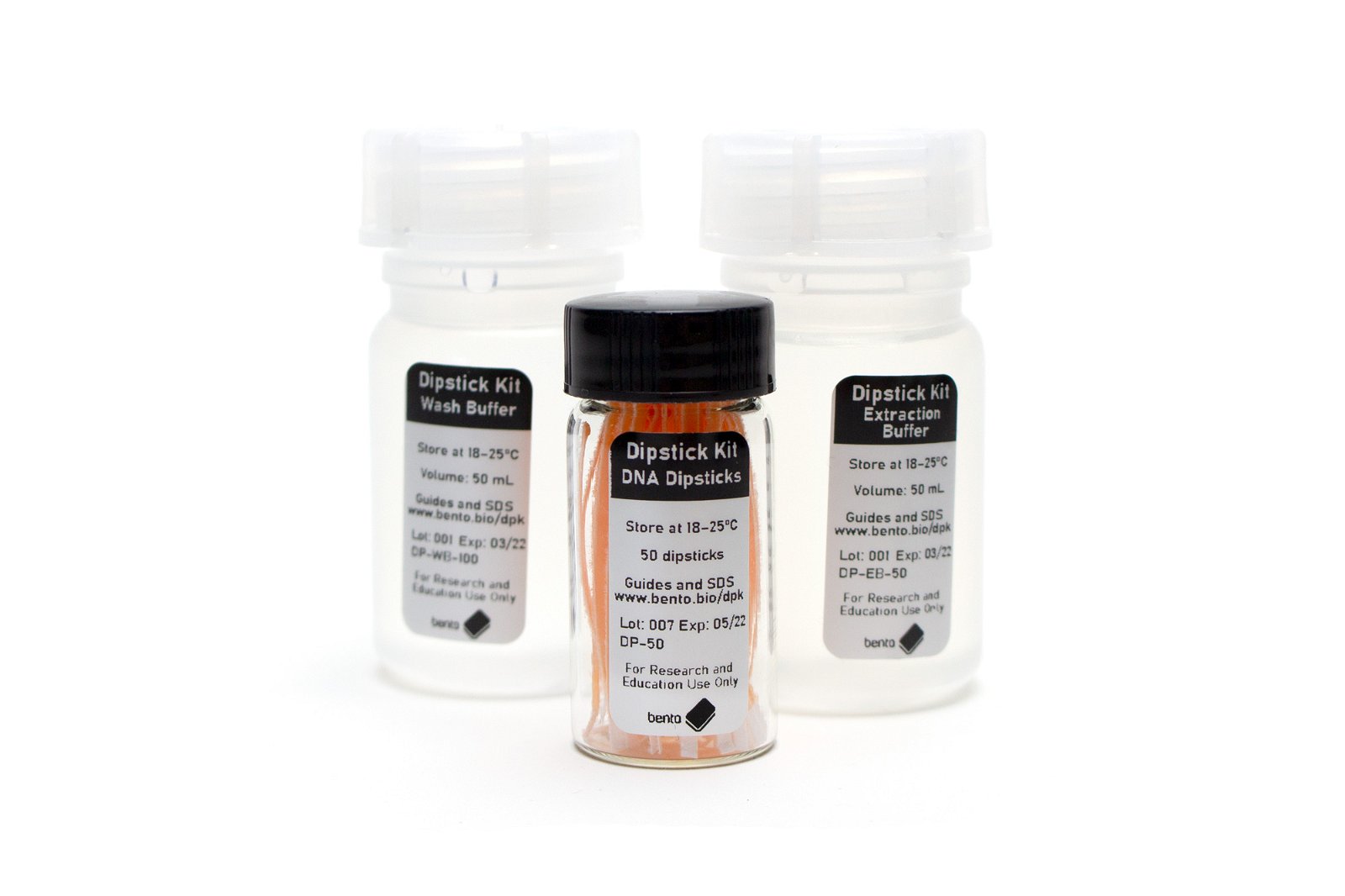
The Dipstick DNA Extraction Method
When to consider the dipstick method
Consider the dipstick DNA extraction method if you need a safe, very quick (<1 min per sample), affordable (<$0.50/sample), equipment-free extraction method to extract a tiny amount of clean DNA that goes straight to a PCR or other amplification mix (LAMP/RPA) or a storage buffer (e.g. TE buffer).
Also, consider dipstick extractions if your samples contain large amounts of PCR inhibitors. The extraction contains a DNA wash step that helps purify the DNA, making it easier to work with challenging samples.
BUT make sure your samples can be ground or disrupted with a micropestle first! If your samples can’t be (e.g. bacterial cells or tough spores, or very hard tissues), then you may need to modify the approach by adding an enzymatic digestion or heating step first.
The dipstick method can work great for DNA barcoding, in-field testing, and teaching environments.
It’s been used for a wide range of different sample types, including bacteria (with enzymatic digestion first), fungi (macrofungi, microfungi, and lichens), invertebrates (crayfish, fish flukes), fish (catfish, salmon), and plants (leaves of many species, including difficult plants like Citrus). It’s a relatively recent technique, and much of its potential is still to be explored!
What does the dipstick method involve?
The dipstick method, developed by Zou et al. (2017) and updated by Mason & Botella (2022), uses a filter paper dipstick to transfer DNA from a crude extraction, through a wash step, into an amplification reaction (PCR, LAMP, RPA). The authors describe three extraction buffers: one based on safe detergents and salts (the version used in the Dipstick DNA Extraction Kit and discussed in the current article) and two using more hazardous chemicals that are not covered here.
The DNA extraction involves a simple four-step process:
- Using a plastic pestle or metal ball bearing, grind or disrupt the sample in a 1.5 mL tube in the extraction solution (a pH-buffered detergent/salt solution containing EDTA).
- Dip a filter paper dipstick (consisting of a small 2-3 mm2 exposed surface and a wax handle) into the extraction solution three (or more) times. A small amount of DNA will bind to the filter paper.
- Dip the dipstick three (or more) times in a wash solution to remove any cellular debris or PCR-inhibiting compounds.
- Dip the dipstick three (or more) times directly into a PCR to release a small amount of DNA suitable for PCR.
Read published studies using our Dipstick DNA Extraction Kit
You can read about some uses of our kit here:
- Leech et al. (2023). DNA-confirmed records of Cortinarius epipurrus and C. hirtus. Field Mycology, 24(3), 83-85. https://doi.org/10.63482/bg9k1e56
- Khajegi et al. (PREPRINT). Rapid Perioperative IDH1 Mutation Detection in High-Grade Gliomas using Novel LAMP assay. https://doi.org/10.21203/rs.3.rs-3207603/v1
- Ruszova et al. (2024). The Utilization of the SaLux19-Based Loop-Mediated Isothermal Amplification (LAMP) Assay for the Rapid and Sensitive Identification of Minute Amounts of a Biological Specimen. Life, 14(5), 579. https://doi.org/10.3390/life14050579
And watch our video here:

Comparing the HotSHOT and Dipstick DNA Extraction Methods
To compare these two methods and see if they meet the criteria for your projects’ needs, consult the table below:
| Criteria | HotSHOT | Dipstick |
| Cost | Very inexpensive, <$0.10/sample (USD) | Inexpensive, <$0.50/sample (USD) |
| Scalability | Very scalable | Not very scalable (each sample must be individually disrupted) |
| Extraction time | Rapid (~30 mins) for all samples (excluding setup and some pipetting) | 30s – 1 min per sample |
| Health and Safety | Safe, but gloves and eye protection are recommended | Safe (but wear gloves anyway) |
| DNA quality | Low (highly fragmented, single-stranded), less suitable for amplicons longer than 500-1000 bp. | Sufficient for PCR, sufficient for 1000 bp amplicons, larger amplicons untested. |
| DNA quantity | Low to moderate | Low |
| DNA purity | Very low, but usually sufficient for PCR with or without dilution | Sufficient for PCR without dilution |
| Sample processing (once in a tube) | Normally none needed | Grinding always needed |
| DNA cleanup step | No DNA cleanup step | Contains a DNA cleanup step |
| Shelf-stability | Stable at room temperature | Stable at room temperature |
| Workflow | Sample → crude extract → neutralised extract → PCR | Sample → crude extract → dipstick → washed dipstick → PCR |
| Suitable applications | Genotyping, DNA barcoding, or detecting most organismal groups | Genotyping, DNA barcoding, or detecting most organismal groups, regardless of PCR inhibitor content |
| It’ll only work if… | • The hot alkaline extraction is enough to release DNA from cells • PCR inhibitors are minimal, or can be diluted below the inhibitory threshold without affecting the PCR | Grinding the sample in the detergent buffer is sufficient to release enough DNA for PCR (otherwise a heat or enzymatic pre-treatment will be needed) |
| Potential modifications | Grinding, microwaving, or freeze/thawing samples before extraction. | • Boiling, freeze/thawing, microwaving, or enzymatic digestion before extraction. • Extracting into TE buffer to create a DNA extract. |
| Unsuitable or unproven applications | • High molecular weight DNA applications • For samples with very high concentrations of PCR inhibitors • Workflows requiring the long-term stability of extracts • Workflows requiring concentration of DNA (e.g.water eDNA) | • High molecular weight DNA applications • Workflows requiring DNA extracts to be stored for future use (although dipsticks can be eluted in TE buffer). • For samples requiring concentration of DNA (e.g. aquatic eDNA) |
Wrapping Up
By showcasing the HotSHOT and dipstick extractions methods, and by making them more accessible via our kits, we hope that more researchers and PCR enthusiasts will be able to access low-cost, rapid, and effective DNA extractions for PCR or LAMP/RPA.
These methods are effective for a wide range of sample and tissue types, provided you don’t use too much material, so they will useful for many different types of project.
And they’re both safe and room-temperature stable, making them useful wherever you might want to use them, whether you’re out on fieldwork; teaching in a classroom; or in a research lab.
But your choice will depend on your project’s needs and priorities. To help you decide, check through the information above to see if they’re a good fit for you.
It would also be worth checking out some of the references below — many of these have useful tips, and they may increase your confidence that these approaches are right for you!
And if you have any questions, please get in touch!
References
HotSHOT DNA extraction method
- Berbegal et al. (2013). A nested-polymerase chain reaction protocol for the detection of Mycosphaerella nawae in persimmon. European journal of plant pathology, 137, 273-281. https://doi.org/10.1007/s10658-013-0237-0
- Bressano et al. (2025). Molecular markers for assisted selection in Sclerotinia blight and peanut smut resistance. Euphytica, 221(3), 1-12. https://doi.org/10.1007/s10681-025-03473-z
- Chan et al. (2024). Eyeing DNA barcoding for species identification of fish larvae. Journal of fish biology, 105(6), 1784-1799. https://doi.org/10.1111/jfb.15920
- da Silvana et al. (2024). An effective DNA extraction method from cajanus cajan seeds suitable for PCR analysis. Bioagro, 36(2), 143-154. http://www.doi.org/10.51372/bioagro362.2
- Elaswad et al. (2021). Genotypic detection of fish-borne zoonotic trematodes using the hotshot DNA extraction method. Egyptian Journal of Aquatic Biology and Fisheries, 25(2), 205-214. https://doi.org/10.21608/ejabf.2021.161826
- García-Abolafio et al. (2023). Simple, fast and inexpensive hot sodium hydroxide and tris DNA extraction method for genotyping tomato and melon seeds. Biotechniques, 75(6), 245-249. https://doi.org/10.2144/btn-2023-0054
- Glover et al. (2016). Genetic screening of farmed Atlantic salmon escapees demonstrates that triploid fish display reduced migration to freshwater. Biological Invasions, 18, 1287-1294. http://doi.org/10.1007/s10530-016-1066-9
- Kagami et al. (2021). Single dominant diatom can host diverse parasitic fungi with different degree of host specificity. Limnology and Oceanography, 66(3), 667-677. https://doi.org/10.1002/lno.11631
- Kinami & Ineno (2025). Alkaline dip DNA extraction from skin mucus for high-throughput sexing of sterlets (Acipenser ruthenus). BioTechniques, 77(2), 56-65. https://doi.org/10.1080/07366205.2025.2467584
- Macgregor et al. (2019). Construction, validation, and application of nocturnal pollen transport networks in an agro‐ecosystem: a comparison using light microscopy and DNA metabarcoding. Ecological Entomology, 44(1), 17-29. https://doi.org/10.1111/een.12674
- Manning et al. (2024). Rapid, equipment-free extraction of DNA from skin biopsies for point-of-care diagnostics. Scientific Reports, 14(1), 13782. https://doi.org/10.1038/s41598-024-64533-3
- Möller et al. (2024). Rich microbial and depolymerising diversity in Antarctic krill gut. Microbiology spectrum, 12(4), e04035-23. https://doi.org/10.1128/spectrum.04035-23
- Montero‐Pau et al. (2008). Application of an inexpensive and high‐throughput genomic DNA extraction method for the molecular ecology of zooplanktonic diapausing eggs. Limnology and Oceanography: Methods, 6(6), 218-222. https://doi.org/10.4319/lom.2008.6.218
- Srivathsan et al. (2021). ONTbarcoder and MinION barcodes aid biodiversity discovery and identification by everyone, for everyone. BMC biology, 19, 1-21. https://doi.org/10.1186/s12915-021-01141-x
- Truett et al. (2000). Preparation of PCR-quality mouse genomic DNA with hot sodium hydroxide and tris (HotSHOT). Biotechniques, 29(1), 52-54. https://doi.org/10.2144/00291bm09
Dipstick DNA extraction method
- Aplin (2021). ‘Sussex Spring Fungus Fortnight’ reveals several new British species and a previously unknown anamorph-teleomorph relationship. Field Mycology, 22(3), 85-90. https://doi.org/10.1016/j.fldmyc.2021.07.006
- Aula et al. (2023). Optimisation of the DNA dipstick as a rapid extraction method for Schistosoma japonicum in infected mice samples and spiked human clinical samples. Infectious Diseases of Poverty, 12(1), 71. https://doi.org/10.1186/s40249-023-01118-8
- Khajegi et al. (PREPRINT). Rapid Perioperative IDH1 Mutation Detection in High-Grade Gliomas using Novel LAMP assay. https://doi.org/10.21203/rs.3.rs-3207603/v1
- Leech et al. (2023). DNA-confirmed records of Cortinarius epipurrus and C. hirtus. Field Mycology, 24(3), 83-85. https://doi.org/10.63482/bg9k1e56
- Mason et al. (2020). An easy‐to‐perform, culture‐free Campylobacter point‐of‐management assay for processing plant applications. Journal of Applied Microbiology, 128(3), 620-629. https://doi.org/10.1111/jam.14509
- Martello et al. (2019). Comparison of the novel dipstick DNA extraction technique with two established techniques for use in biological barcoding. Molecular Biology Reports, 46, 6625-6628. https://doi.org/10.1007/s11033-019-05083-0
- Ruszova et al. (2024). The Utilization of the SaLux19-Based Loop-Mediated Isothermal Amplification (LAMP) Assay for the Rapid and Sensitive Identification of Minute Amounts of a Biological Specimen. Life, 14(5), 579. https://doi.org/10.3390/life14050579
- Zou et al. (2017). Nucleic acid purification from plants, animals and microbes in under 30 seconds. PLoS biology, 15(11), e2003916. https://doi.org/10.1371/journal.pbio.2003916
Please let us know what other resources, advice, and tips and tricks for using Bento Lab that you would like us to produce in the future!
Looking for advice on using Bento Lab?
Book a free consultation or ask a question.